
Modern diagnostics and advanced urological treatment
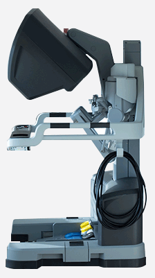
Medicover Hospital is one of three medical centres in Poland that owns the most advanced robotic system in the world. Surgeries assisted by da Vinci robot are characterised by exceptional precision and low invasiveness.
Using the da Vinci robot increases the effectiveness of surgery and significantly reduces the risk of complications.
One of the leading experts in Europe using this system is Md PHD Paweł Salwa, a urological surgeon. With the help of da Vinci robot, Md PHD Paweł Salwa has already operated on over 2000 patients and based on this extensive experience he has created an original surgical method “SMART Prostatectomy”, which is now also available to Polish patients.
da Vinci®
is the art of treatment
All urological procedures are carried out by specialists with many years of experience.
In emergencies, we offer urgent medical help, including emergency surgical procedures.


LEARN MORE
MODERN DIAGNOSTICS
In most cases, prostate cancer in its early stages is asymptomatic, and the reason for urological consultation is an abnormal level of the prostate-specific antigen (PSA) in the blood.
The European Association of Urology (EAU) recommends that men aged 50 (or 45, if prostate cancer runs in the family) undergo regular urological check-ups and the PSA blood test.
If the PSA level exceeds 4 ng/ml, further diagnostics should be performed.
Current European guidelines recommend undergoing at least a 12-core prostate biopsy through the rectum. A standard transrectal biopsy using a transrectal ultrasound (TRUS) may not detect small cancer foci, not visible through this method, or inaccessible due to their location.
MRI OF THE PROSTATE
The solution to this problem is to perform a high-quality imaging test that allows you to locate the change and then collecta biopsy sample from the changed area. This test is the multiparametric magnetic resonance imaging of the prostate (mpMRI).
It has been shown that the sensitivity (the ability to detect cancer) of the prostate magnetic resonance is almost twice as high (72% compared to 38%) of aregular TRUS.
However, the urologist’s experience is essential here as he will interpret these images and use this information to properly perform the targeted fusion biopsy.
PROSTATE FUSION BIOPSY
At Medicover Hospital in Warsaw, the prostate fusion biopsy is performed under general anaesthesia (without the pain or discomfort that usually accompanies a regular biopsy) using Medcom’s advanced technology. This biopsy takes place through the groin, which eliminates the risk of prostate inflammation or sepsis, which is a risk for patients undergoing a standard transrectal biopsy.
Patients are well enough to return home the day after the procedure.
Further treatment depends on the result of the histopathological examination.
If prostate cancer is detected and surgery is required, it can be performed with the best method available for patients, which is the da Vinci robot.
If the biopsy results exclude the presence of prostate cancer, we can then focus on effectively treating the patient’s symptoms.
Patients are qualified for the fusion biopsy through the prostate magnetic resonance during a urological consultation at Medicover Hospital.
The prostate MRI is performed at Medicover Hospital’s Magnetic Resonance Laboratory before the planned fusion biopsy.
LEARN MORE
Prostate cancer treatment with the latest Da Vinci Robot
Using the da Vinci robot increases the effectiveness of surgery and significantly reduces the risk of complications. One of the leading experts in Europe using this system is Dr Paweł Salwa, a urological surgeon currently working at Medicover Hospital, and formerly the Head of the largest Robotic Urology Clinic in Gronau, Germany. With the help of the da Vinci robot, Dr Salwa has already operated on over 2000 patients and based on this extensive experience he has developedan original surgical method “SMART Prostatectomy”, which is now also available to Polish patients.
Radical prostatectomy assisted by da Vinci robot is a minimally invasive method of prostate cancer treatment, consisting of completely removing the cancerous prostate gland. The procedure is performed under general anaesthesia. Several 1.5 cm holes are cut in the abdomen through which four robotic arms are inserted into the patient’s body. A stereoscopic camera is placed on one of the arms, allowing to obtain a three-dimensional image of the operating field. The remaining arms are equipped with miniature surgical instruments, allowing for precise handling of individual tissue fibres.
Radical prostatectomy assisted by da Vinci robot is the most modern and technologically advanced method of prostate cancer treatment. The reason why urologists around the world are so keen to use this technology is because it allows for a very precise surgical procedure. Tenfold enlargement of the image, full HD, tools measuring 5 mm, and the ability to move in any direction the operator wishes, all allow to obtain the most important objective–to completely remove the tumour. The precision of the procedure, apart from the oncological result, has one more aspect – it helps maintain potency and control over urinary retention.
LEARN MORE
Bladder cancer
The presence of blood in the urine is an alarming symptom that should not be ignored because up to 20-25% of patients with haematuria have bladder cancer.
Therefore, every, even a single (self-limiting) episode of haematuria requires proper urological diagnostics to confirm or rule out urinary tract cancer.
The main risk factor for bladder cancer is long-term smoking. Bladder cancer most often occurs after the age of 60. In Poland, men are three times more likely to be diagnosed with bladder cancer than women.
Bladder cancer diagnostics
Cystoscopy is a test involving inserting a special instrument into the urinary bladder through the urethra, allowing a precise assessment of its interior. The examination can be performed using a flexible cystoscope at the urological clinic (under local anaesthesia). If the urologist finds abnormal changes during the cystoscopy, it is advisable to remove them in the hospital conditions and perform a histopathological assessment.
If imaging examinations, such as an ultrasound of the abdominal cavity, revealed a bladder tumour, most often cystoscopy is performed in the operating theatre under general or regional anaesthesia –in this case, apart from visual assessment, it is possible to remove the tumour using a minimally invasive method. This procedure is called transurethral resection of a bladder tumour (TURBT) After the procedure, a catheter is left in the urinary bladder for approximately 24 hours. Most often, patients can go home the day after surgery. The results of the histopathological examination are available after about 2-3 weeks.
Further urological procedures depend on the severity of the cancer. Bladder tumours infiltrating the deep wall of the bladder (infiltrating the muscle membrane) require radical removal (cystectomy). Prior to that, additional imaging examinations (computed tomography or magnetic resonance) are performed to assess the severity of the disease. If you qualify for surgery, a radical cystectomy may be performed with the help of a da Vinci robot.
Cancers of the upper urinary tract
Cancers of the upper urinary tract (renal pelvis and ureter) are much less frequent than bladder cancer – they constitute 5 to 10% of all malignant urinary tract cancers.
They occur most often in people over 70 years old, three times more often in men. The most important risk factor for the development of upper urinary tract cancer (as in the case of the bladder) is chronic smoking. In the majority of patients, the only sign of the disease is haematuria. Additional symptoms such as pain in the side, weight loss, and fatigue may occur in more advanced cancers. Diagnosis usually starts with an abdominal ultrasound. The examination confirming the diagnosis is abdominal and pelvic tomography with contrast with urographic phase (alternatively magnetic resonance with contrast). Control cystoscopy is also recommended to exclude co-existing bladder cancer, and urine cytology should be performed.
The scope of treatment depends on the location and severity of the disease. In the case of high-risk urinary tract cancers, surgery is recommended. The surgery involves removing the kidney along with the entire ureter (nephroureterectomy). At Medicover Hospital in Warsaw, this treatment is performed by the da Vinci robot.
LEARN MORE
Malignant kidney diseases – kidney cancer
Kidney cancer is usually asymptomatic in its early stages and is often detected accidentally during an imaging check-up (most commonly an abdominal ultrasound) or during the diagnosis of other diseases. The proven risk factors for kidney cancer are obesity, hypertension and smoking. If an abdominal ultrasound reveals suspected kidney cancer, the next step is to perform abdominal cavity examinations with contrast (computed tomography or magnetic resonance). If on the basis of the diagnosis we suspect kidney cancer limited to the organ, the patient is qualified for surgical treatment, the scope of which (removal of the tumour or removal of the entire kidney) depends on the severity of the disease.
At Medicover Hospital in Warsaw, we have a full range of methods for minimally invasive surgical treatment of renal tumours –surgery assisted by the da Vinci robot, as well as the laparoscopic method. The method and scope of surgery is tailored individually to the patient’s needs after prior urological consultation.
KIDNEY CYSTS
Kidney cysts are fluid spaces located in the parenchyma. They are the most common focal lesions of the kidneys. In most cases, they are asymptomatic and detected accidentally during an abdominal ultrasound. Very large cysts (> 10 cm) may be accompanied by pain in the lumbosacral region resulting from pressure on the neighbouring structures. During diagnostics, kidney cysts should be differentiated between simple (most frequent, not cancerous) and other types of cysts, including lithocysts that may point to kidney cancer. Suspected cancerous lesions are verified by performing an abdominal cavity contrast study (computed tomography or magnetic resonance).
Further steps depend on the nature of the cyst – its size, morphology, and accompanying symptoms. In the case of symptomatic simple cysts, we perform laparoscopic cyst removal. Cystic lesions with a high risk of malignant cancer are treated surgically as solid tumours – surgery with da Vinci robot or laparoscopy is possible. The method and scope of surgery is tailored individually to the patient’s needs after prior urological consultation.
THE URETEROPELVIC JUNCTION (UPJ) STENOSIS
Stenosis of the ureteropelvic junction is a congenital or acquired disorder of the upper urinary tract causing difficulties in urine flow from the kidney, accompanied by an increase in pressure above the blockage. This condition may lead to a progressive atrophy of the renal parenchyma and its insufficiency, recurrent infections or the formation of kidney stones.
The most common symptoms are pain in the lumbar region, which may be more severe after drinking a large amount of fluids. They may be accompanied by nausea, vomiting, sometimes fever (in case of infection). The disease may be asymptomatic and may be diagnosed during an ultrasound examination of the abdominal cavity.
Imaging tests (abdominal ultrasound, urography, tomography with urographic phase),as well as examinations assessing renal function (renoscintigraphy) are used in the diagnostics and treatment of the narrowing of the ureteropelvic junction.
Surgery is recommended in the case of a symptomatic form of the narrowing and/or progressive impairment of kidney function. The standard surgery performed is pyeloplasty, which can be performed using the da Vinci surgical robot.
LEARN MORE
PHIMOSIS
It is a condition in which the foreskin cannot be pulled back from the glans due to its narrow, often scarred opening. The surgical treatment consists of removing the foreskin (circumcision) or plastic surgery of the foreskin leading to the widening of its opening. This procedure may be performed under short-term intravenous or local anaesthesia,which allows the patient to leave the hospital on the same day.
SHORT FRENULUM
A short frenulum is accompanied by pain and discomfort during pulling back of the foreskin off the glans, due to the short frenulum (skin fold, located on the underside of the glans, between the glans and the foreskin). These symptoms intensify during an erection, making intercourse difficult.
Treatment involves plastic surgery of the frenulum –which is performed within the One Day Surgery Unit.
VARICOCELE
One of the main causes of male infertility is varicocele, which accounts for 40% of cases of this disease. It affects about 20% of men, but many of them only learn of the diagnosis after many unsuccessful attempts to have a child. Others learn about varicocele when symptoms such as heaviness in the testicles and groin area become very painful, and the varicose veins become noticeable.
In each case, however, the problem is serious, because varicose veins obstruct the outflow of venous blood from the testicles, increase their temperature and the pressure of fluids contained in them, and as a result lead to the disturbance of sperm morphology, its quality and sperm life. Fortunately, early detected,
varicocele may be cured, and the infertility it causes is reversible. However, it requires the help of a specialist and excellent conditions to perform the surgery.
The basic diagnostic method for suspected varicocele is a non-invasive ultrasound examination, which clearly shows the disturbances of the flow of venous blood in the testes and the deformation of blood vessels. The ultrasound allows for a quick and accurate diagnosis, defining the stage of disease, and thus allowing to choose the most appropriate method of treatment:
- laparoscopically: a particularly effective method for young men with no anatomical disproportions in the size of the testicles. The minimally invasive procedure under general anaesthesia is safe and complication-free. The laparoscopy leaves only three small incisions. The recovery period lasts up to 14 days.
- operationally (“classic method”): an invasive method involvingmore complications. Recommended for elderly patients and those with advanced (II and III) stages of the disease. It most often involves the retroperitoneal ligation of the testicular vein in its middle section. Stitches are removed 10 days after the surgery and the recovery period lasts 30 days.
HYDROCELE
A hydrocele is the accumulation of fluidsaround the testicle. It may be the result of injury, surgery, other procedure, or an inflammation that develops within the scrotum. Sometimes it can be a symptom of testicular cancer. Long-lasting hydrocele may adversely affect sperm production. In its advanced form, it may even lead to infertility and atrophy of the testicle. The procedure is performed within the One Day Surgery Unit.
SPERMATOCELE
The spermatocele is located in the scrotum near the testis or epididymis, where thesperm cells occur. Sometimes it causes pain, making it difficult to perform daily tasks. The cyst is removed during surgery withinthe One Day Surgery Unit.
LEARN MORE
CONSULTATION IN THE FIELD OF
cancer and other prostate diseases, as well as da Vinci surgery
Paweł Salwa MD – Head of Urology Department
To book a consultation please call: +48 794 407 279
UROLOGICAL CONSULTATIONS
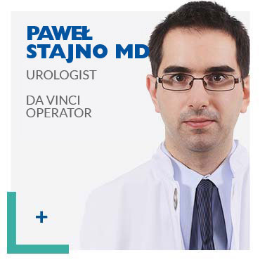
Paweł Stajno MD
Marcin Rudziński MD PhD
Piotr Dzigowski MD
To book a consultation please call: +48 500 900 900

LEARN MORE
Paweł Salwa Md PHD
Head Of Urology Department At Medicover Hospital
PROSTATIC CANCER CONSULTATIONS
Md PHD Paweł Salwa took over as Head of the Urology Department at Warsaw’s Medicover Hospital in early 2019. Prior to that, hehad worked in the largest robotics clinic in Gronau, Germany for 7 years. Since 2013, he has been treating prostate cancer using the minimally invasive da Vinci robotic method.
He has since performed over 2000 operations using the da Vinci robot. As a leader in implementing new surgical methods, he has developed the original SMART Prostatectomy method, which allows him to achieve very good results.
For Md PHD Paweł Salwa, the goal of every surgery is:
- complete removal of the tumour
- full continence control
- return to sexual activity
LEARN MORE
Paweł Stajno Md
Urologist At The Urology Department At Medicover Hospital, da Vinci Operator
UROLOGICAL CONSULTATIONS
Paweł Stajno is a graduate of the Medical University of Warsaw, graduating with distinction in 2010. During his studies, together with Dr Salwa, he had won university and nationwide individual and team competitions of anatomical and biochemical knowledge.
He has received multiple scholarships for outstanding academic results. He completed his specialisation in the field of urology at the Urinary Tract Cancer Clinic of the Cancer Centre in Warsaw, receiving the prestigious FEBU title (Fellow of the European Board of Urology), obtaining one of the five best results among those taking the EBU exam in Poland. Paweł Stajno is a member of the Polish and the European Urological Association.
Dr Stajno constantly improves his knowledge and skills by participating in conferences and trainings. He is the author and co-author of numerous publications, including chapters in urological monographs. Doctor Paweł Stajno has been a member of the urological team at the Polish Robotic Urology Centre at Medicover Hospital from the beginning of its operation, dedicating his time to treat patients at the Centre. Dr Stajno specialises in the diagnosis and treatment of urological diseases, with particular emphasis on uro-oncology and functional urology. He speaks fluent English.
LEARN MORE
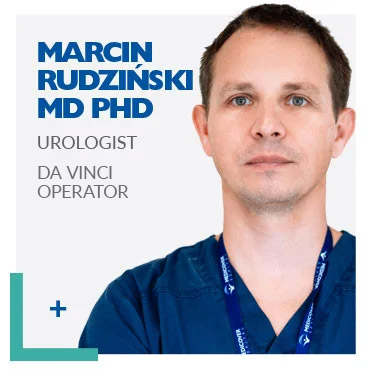
Marcin Rudziński MD PhD
Urologist At The Urology Department At Medicover Hospital, da Vinci Operator
Urology specialist, FEBU (Fellow of the European Board of Urology), member of the Polish Society of Urology and the European Association of Urology since 2013.
Graduate of the First Faculty of Medicine of the Medical Academy in Warsaw in 2005.
Gained professional experience in the General and Vascular Surgery Departments of the Bielański Hospital and the Urology Clinic of the Postgraduate Medical Education Center at the Professor W. Orłowski Hospital in Warsaw.
From 2013 to 2020 he worked as a Senior Assistant at the Urology Department of the Międzylesie Specialist Hospital in Warsaw.
In March 2017 he obtained the title of doctor of medical sciences, defending his dissertation on the treatment of urolithiasis in patients with short bowel syndrome.
Specialises in oncological and functional urology.
An experienced, committed professional with an individual approach to the patient.
LEARN MORE
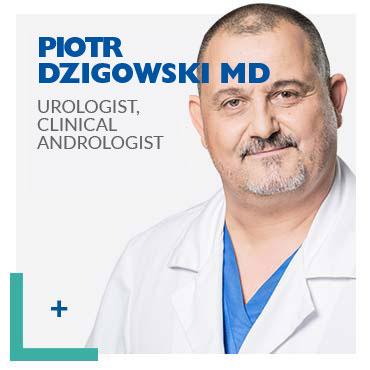
dr Piotr Dzigowski
Urologist, Clinical Andrologist
A graduate of the Faculty of Medicine at the Collegium Medicum of the Jagiellonian University (1997). In 2005, he became an Assistant at the Department of Urology at the Medical University of Warsaw, and in 2010 he obtained his specialisation in urology and membership in the FEBU
(Fellow of the European Board of Urology). He is an expert in general, oncological and functional urology.
Dr Dzigowski deals with the diagnosis and treatment of urolithiasis, urinary incontinence, neurogenic bladder disorders, benign prostatic hyperplasia, urinary tract infections and varicocele. In addition, he has many years of experience in treating patients with urinary tract tumours – cancers of the kidneys, ureters, bladder and prostate, as well as the urethra, penis and testicles.


You can reach us by public transport by buses 217 or 522. The bus stop (Branickiego) is located approx. 50 metres from the hospital. GPS: 52,15513 N, 21,07863 E, 52’9’18 N, 21’4’43 E
Dear Patients,
we would like to inform you that the three-level car park located opposite the entrance to the Hospital has narrow passages. Therefore, it can be difficult to manoeuvre (especially larger vehicles). In this situation, we recommend to use the external car park, which has 117 parking spaces and is located at the back of the building.


Contact
Appointment booking
Telephone: +48 794 407 279
Address
Medicover Hospital
al. Rzeczypospolitej 5
02-972 Warszawa
(WilanówTown)
Copyright © Medicover 2019


